Surgical robot takes shape
Five years ago, the MIRACLE team at the University of Basel began their work on revolutionising the future of bone surgery. Now the research team – which is financed by the Werner Siemens Foundation – have put the various components of their unique system together for the first time and presented it to the media.
Bone surgery can be a brutal, bloody affair. Conventional bone cutters and drills lack precision and require enormous amounts of force to function – secondary damage to the surrounding tissue is often the unpleasant consequence. Researchers in the MIRACLE project at the University of Basel are aiming to transform bone surgery by developing a revolutionary system to make interventions safer, faster and more precise. It’s an ambitious goal that won’t be achieved overnight. “You could say we’re the Mars mission in the field of medical robotics research,” says Philippe Cattin, one of four project leaders in MIRACLE.
Now, after five years of research, the team have reached a major milestone. At a media conference in early September, the researchers presented a complete system in which their separate, newly developed technologies were integrated to form a modular robot that will one day make minimally invasive bone surgery a reality.
For that to happen, Cattin says four state-of-the-art innovations must be perfected. He and his team have already developed the first: a virtual and augmented reality system to aid surgeons in planning and monitoring operations. The tool generates three-dimensional, animated models of the body part to be operated on. In this virtual space, doctors can peer into a patient’s body and view a fracture or a tumour from all angles. The system is already a standard procedure in several types of surgical interventions at University Hospital Basel.
Tiny endoscope
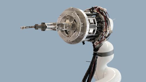
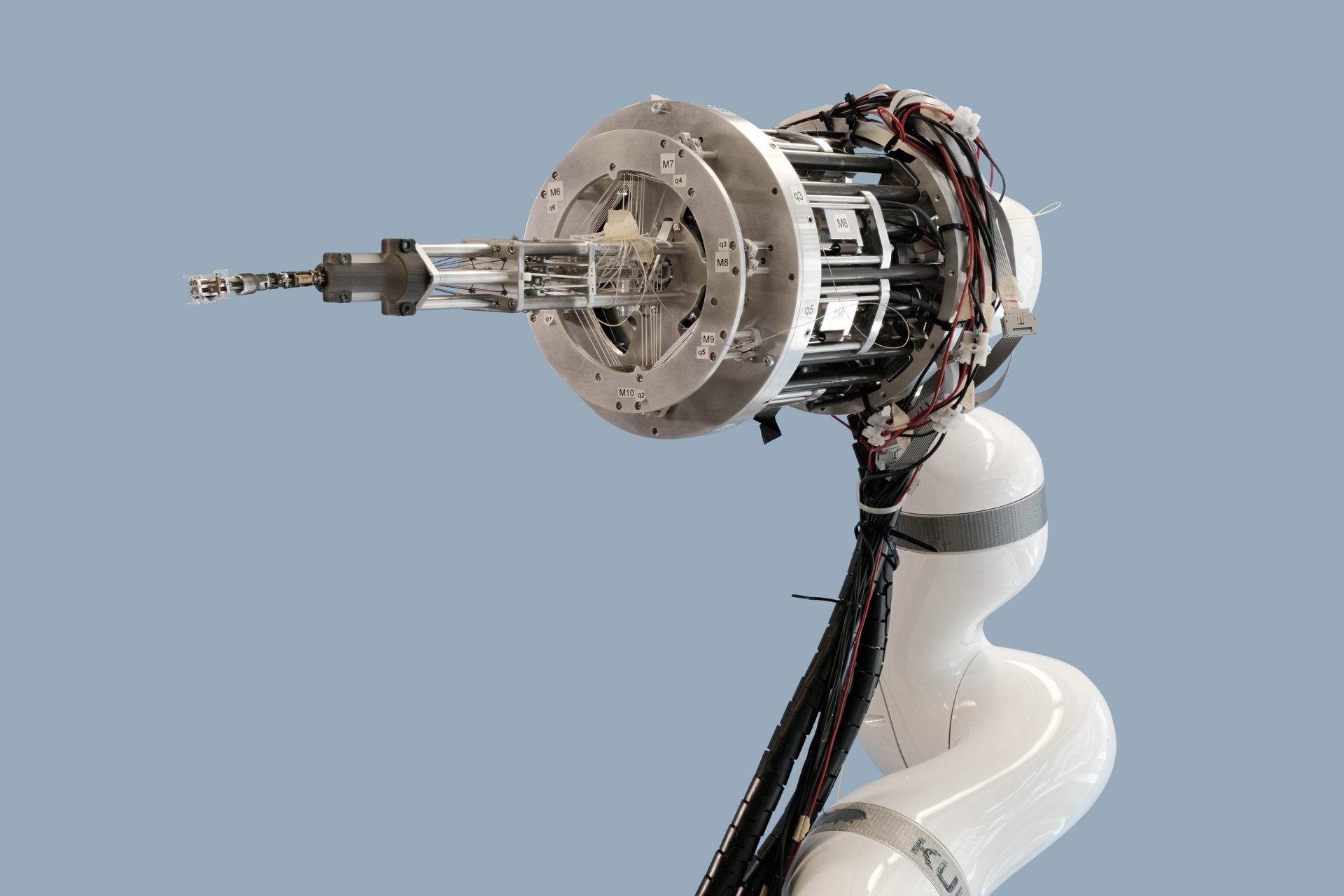
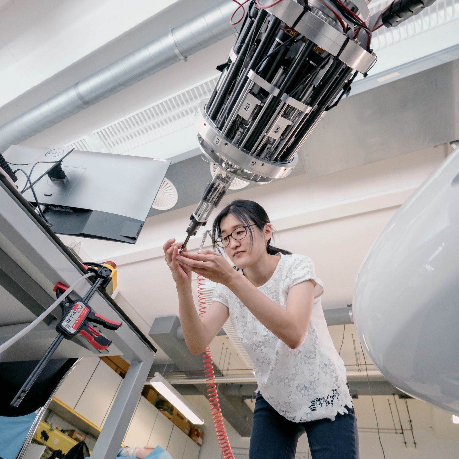
The second innovation is being designed in the lab of Georg Rauter, mechatronics specialist, engineer and head of the robotics group in the MIRACLE project. He and his team are developing a robot that can insert a tiny endoscope in the body, steer it to the correct position and actuate several instruments there. Built to function akin to a human hand, the robotic “arm” with its “hand” will be mounted on the ceiling of the operating theatre, from where it will navigate its finger-shaped extension containing the endoscope to the surgical site on the patient’s body.
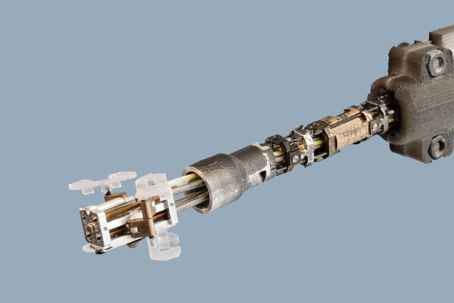
One of the major challenges facing the team is the navigation system: when taking over control, it’s important that a surgeon’s movements can be intuitive. To solve this problem, Rauter and his team have come up with a clever method that predicts where a doctor’s hand will move based on the pressure it exerts. Another challenge is miniaturisation. Because the foremost part of the robot, which penetrates down to the bone, should be no larger than half a centimetre, the actuator system must be located outside the patient’s body. These “motors” are connected to the instruments at the tip of the robot – the finger, as it were – via a tendon-like material. “Minimally invasive interventions would otherwise be impossible,” Rauter explains.
Precise incisions
The key component of the future robotic surgeon is a laser that functions like a scalpel. Unlike conventional bone cutters, the laser scalpel can be made so small that it can be used in minimally invasive interventions. A further advantage is that it makes much cleaner, more precise incisions. This means the bone remains intact where it’s been cut, “as though it doesn’t notice what’s happened to it,” says Ferda Canbaz, head of MIRACLE’s laser lab. These clean incisions heal much more quickly than the cuts made with today’s instruments, which can badly damage the edges of a bone.
To prevent the laser from cutting the wrong tissue, it’s critical that the surgeon always knows the laser’s exact position, and developing this kind of “smart” laser is the third innovation in the project. Canbaz and her team are exploring two approaches: the first works with spectroscopy, a method that uses plasma light to determine the type of tissue being cut. The other approach is an optical system capable of measuring the depth and form of an incision as well as body temperature at the operation site.
Made-to-measure implants
Another major advantage the laser scalpel has over conventional instruments is that surgeons can cut a variety of shapes with it – for instance, shapes into which an implant can be inserted like a missing puzzle piece. This is the starting point for Florian Thieringer, maxillofacial surgeon at the University Hospital Basel. His work focuses on the fourth innovation in the project: 3D printed implants. At the hospital’s inhouse 3D Print Lab, Thieringer plans to produce implants that fit seamlessly into pre-cut bones and that stay in place without being affixed with screws or plates, as is currently the practice.
To ensure the implants are small enough to use in minimally invasive procedures, Thieringer is experimenting with a type of origami technique. His idea is to make implants consisting of numerous small parts that the surgeon inserts into the patient’s body, one at a time, and then assembles in vivo.
The MIRACLE mission will need a few more years before it’s “ready to orbit Mars”. But once the system has been perfected, patients will no longer have large wounds and scars, they’ll be able to put weight on their bones soon after surgery and will hardly notice the implant in their body. All thanks to four innovations made in Basel.
