A portable heartbeat
Placing an artificial muscle in the aorta to support the hearts of patients with cardiac insufficiency—this is the aim pursued by researchers at the Center for Artificial Muscles. And they are nearing their goal in leaps and bounds: after just a three-year development phase, they have now tested the first prototype of their artificial muscle on a pig heart—with very encouraging results.
Some two hundred thousand people in Switzerland suffer from cardiac insufficiency. Medication is generally prescribed to treat the disease, but the heart’s pumping capacity often diminishes over time. In severe cases, ventricular pumps are inserted to help the weakened organ do its job until a donor heart is available. However, this life-saving measure has its price: invasive surgery is necessary to place the mechanical pumps inside the heart, where they can cause wear and tear on the heart lining and destroy red blood cells. Now, researchers at the Center for Artificial Muscles (CAM) at the École polytechnique fédérale de Lausanne (EPFL) are working on a less invasive solution—an artificial muscle that is fitted into the aorta to give weak hearts a boost.
Soft and elastic
In contrast to ventricular pumps, the artificial muscle consists of a soft, elastic material and does entirely without rigid metal parts. “It’s malleable and can meld with the tissue,” says microengineer and CAM director Yves Perriard. The muscle expands and contracts to change the circumference of the aorta and thus the pressure exerted on the left ventricle of the heart, which is responsible for pumping oxygenated blood through the aorta and into the body’s circulatory system. The principle is as follows: if the artificial muscle helps the aorta dilate at the right time, the left ventricle can do its job of pumping blood through the body with less effort.
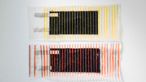
In essence, the artificial muscle is a plastic membrane encased in carbon layers capable of conducting electricity—and electrical impulses are what control the membrane. When an electrical current is switched on, the membrane expands; when the voltage is switched off, the membrane contracts again. The prototype membrane is encased in a stabilising plastic scaffold and covered with an additional thin layer to seal it off from body fluids.
Lab experiments were conducted and the principle was proven to work: to test the artificial muscle, the team developed an apparatus that simulates both heart chambers as well as blood circulation through the aorta. The muscle functioned as planned and reduced pressure in the left ventricle.
First operation a success
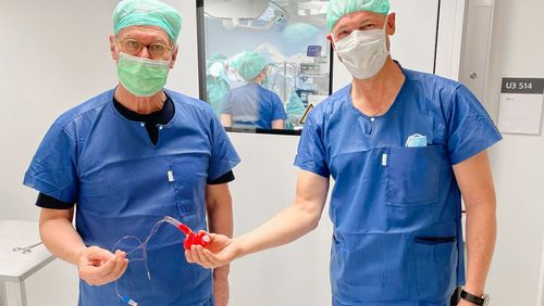
The first animal experiment was carried out in April of 2021. In a complex operation, surgeons made an incision in the aorta of a pig and inserted the artificial muscle, while also positioning several sensors that measure how the aorta muscle impacts the heart and circulation. “This trial was an extremely important milestone,” says Yoan Civet, Managing Director of CAM. “If the aorta ring hadn’t worked in the animal experiment, we would have had to completely rethink our approach.”
But the test was a success—to put it mildly. The pressure on the animal’s left ventricle dropped, and the impulses from the aorta ring helped the pig heart pump more efficiently. The heart muscles also expended less energy, and the flow of blood through the aorta increased. Moreover, the researchers gained a detailed understanding of how the movements of the artificial muscle in the aorta should be aligned with the pulse to provide optimum support to the heart.
Pocket-sized voltage
Currently, the artificial muscle can boost the pumping capacity of the left ventricle by roughly ten percent. About twice as much would be needed to support a human heart in the long term. “We’re now focusing on finding ways to convey the necessary ten thousand volts into the body and to the artificial muscle,” Perriard says. At present, electric cables are used to conduct the voltage; in future, however, the aim is to develop a wireless, portable power source. But “generating this much voltage in a small device is a major technical challenge”, says Perriard. The team aim to miniaturise the electronic power supply in a step-by-step process by first reducing it to the size of a small box measuring roughly twenty-by-ten-by-ten centimetres, then to the dimensions of a device so small that patients could wear it.
The researchers also have plans to take the design of the aorta ring to the next level. Specifically, they are seeking a way to wrap it around the aorta, thus avoiding an incision and preventing the artificial muscle from coming into contact with blood.
The heart is just a start
Artificial muscles could also help patients with facial paralysis or people suffering from urinary incontinence. The team have already launched their research into these areas. To alleviate urinary incontinence, the researchers aim to design a small ring that would be fitted around the urethra to function as an additional sphincter. For use in facial surgery, the team are collaborating with the University Hospital Zurich to develop flat membranes able to support, and possibly even replace, facial muscles.
Text: Santina Russo
Photo: Center for Artificial Muscles (CAM)
 to squeeze and then relax the urethra.")