Observing the living cell
Despite major advances in modern medicine, there are still many diseases that evade detection and treatment. To remedy this, researchers at the Werner Siemens Imaging Center in Tübingen are developing new imaging techniques that enable greater precision when examining tissues and molecules. Recently, the team have made great progress in cancer therapies and the early detection of Parkinson’s disease.
Cancer is the leading cause of early death in the Western world—and in Germany and Switzerland, most deaths in men over forty-five and in women over twenty-five are cancer related. “Although we have good treatments for many cancers, the disease often returns after a few years,” says Bettina Weigelin, group leader at the Werner Siemens Imaging Center (WSIC) in Tübingen, Germany. With her team, she is seeking methods to better characterise tumours and to improve cancer therapies with this new-found knowledge.
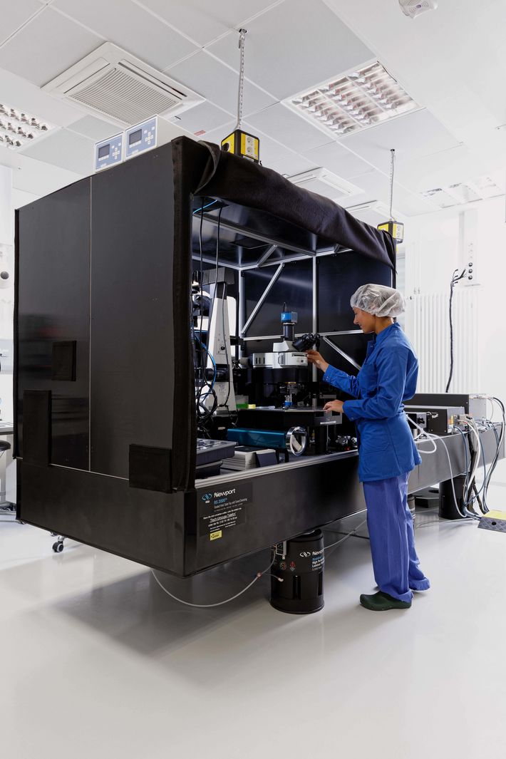
To achieve their ambitious goal, the Tübingen researchers have a powerful new tool: their intravital microscope, a microscopy technique used to examine living tissue, even entire organisms. The microscopy system, which was set up at the start of 2021, fills an entire room—a lab space where researchers are required to wear disposable overalls. In addition to the microscope itself, the system has three high-energy infrared lasers. “With this laser technology, we can actually see inside a sample, not just observe its surface,” Weigelin explains.
To examine a specimen, the researchers first mark the cells they want to track—often tumour and immune cells—with fluorescent proteins. Under the microscope, the lasers then stimulate the cells, rendering them visible down to a tissue depth of two millimetres. “Only very few intravital microscopes in the world can achieve this depth penetration,” says Weigelin.

In the lung of a mouse
Weigelin and her team are mainly interested in better understanding how immunotherapies influence tumours. They are compiling this knowledge through experiments on mice in which the animals are given a mild anaesthetic and placed under the microscope. Previously, the researchers had focused on skin cancer, but the new unit allows them to analyse tumours that are situated deeper in the body. They are now preparing their first breast cancer experiments.
The team have also begun investigating metastases. “With many types of cancer today, the primary tumours are no longer the cause of death. Rather, patients die of metastases in their bones or internal organs,” says Weigelin. On her computer, she opens images of a mouse’s lung, where several round spots are glowing: metastases. These images were made using another kind of microscopy—light sheet fluorescence microscopy. Although this technique does not render live organisms visible, it illuminates layers, or slices, of entire organs. The magnified image of the metastases in the mouse’s lung reveals how the immune cells are fighting the tumour. At the edge of the growth, however, it becomes apparent that the dividing tumour cells are winning the battle. “Although the immune cells often recognise the tumour cells, they’re unable to destroy them,” says Weigelin. “We want to find out why this is the case and use this understanding to develop new therapies.”

Destroying sleeper cells
The WSIC group led by the centre’s director, Professor Bernd Pichler, is studying another phenomenon in tumour cells: senescence. Cells in a senescent state are still alive, but they no longer divide; several cancer drugs exploit this mechanism to stop the growth of tumours. However, as Pichler explains, “it’s impossible to drive all of a tumour’s cells into a senescent state”. Moreover, senescent cells secrete signals that stimulate growth in the other cancer cells. “As a result, tumours that had been suppressed begin to grow again,” Pichler says. “To prevent this from happening, we need to pinpoint exactly when we should destroy the senescent cells with drugs.” To determine this precise moment, the team have developed a senescence tracer, which consists of a molecule that marks senescent cells with radioactive tracers, making the cells visible in a positron emission tomography (PET) scan. The researchers at WSIC have already tested their method in preclinical trials. Currently, a phase I clinical trial is under way—the world’s first-ever clinical trial of a senescence tracer.
The method has already delivered encouraging results. In collaboration with the University Hospital Tübingen, Pichler’s team used the senescence tracer on patients with glioblastoma tumours that had proved resistant to all other therapies. Although these brain tumours are highly aggressive, patients whose treatments were administered on the basis of a PET scan with the senescence tracer lived longer. “One fifty-year-old patient lived several more months with a good quality of life,” says Pichler.
 and her team can use the new intravital microscope to precisely examine the influence of immunotherapy on tumors. Even tumors deeper in the body, such as breast cancer, can be analyzed.")
Early detection of Parkinson’s
The WSIC team are also researching early detection of neurodegenerative diseases like Parkinson’s. “In Parkinson’s, a certain type of protein, so-called Lewy bodies, are deposited in the brain long before the first symptoms appear,” says group leader Kristina Herfert. She and her team have now developed the world’s first tracer that binds to Lewy bodies, rendering them visible in the PET scan. “This delivers a reading that shows how the disease has progressed thus far,” explains Herfert. In future, it is hoped that the new tracer will help doctors to monitor whether a therapy is working—perhaps even make early detection of Parkinson’s a reality. After promising results in preclinical trials with mice and with human brain tissue, the plan is to test the tracer already next year in a clinical trial with patients.
Recently, WSIC group leaders Kristina Herfert and Bettina Weigelin were appointed professors at the University of Tübingen, prevailing over formidable international competition in the appointment procedure. “It’s evidence of how strong our team is, and it also enhances the reputation of our centre,” says Pichler. “I’m very proud.”
Text: Santina Russo
Photos: Oliver Lang
 is an international leader in the development of low-level radioactive tracers used in medical imaging.")