

Growing skin in a Petri dish
For more than a quarter century, clinicians and researchers at the University Children’s Hospital Zurich have been developing a skin substitute to treat children and adolescents with severe burns. They’ve now further refined their innovation, making it even more like real skin. Thanks to funding from the Werner Siemens Foundation (WSS), the first clinical trials with patients can proceed.
Touching a hot stove or baking tray, playing with matches, an accident with a deep-fryer, a knocked-over teakettle: burns and scalding injuries happen quickly—especially when children are involved. In most cases, the wounds are superficial and heal quickly, but in others, the skin is so severely damaged that medical care is needed. In 2019, more than nine million people worldwide sustained burns or scalding; of these, 111,000 died from their injuries and more than 1.5 million were left with lifelong disabilities.
These types of injuries are treated at Switzerland’s only centre for paediatric burn victims, located at the University Children’s Hospital Zurich. However, the centre offers more than just burn treatments. Clinicians and researchers are also developing a therapy that has the potential to drastically improve the lives of burn patients—the world’s first skin substitute that is very similar to real skin. Their product, PV-Skin (P=Pigmented, V=Vascularised) has now progressed to the stage where it’s ready to be tested on patients. The next step is to control its safety in humans, and over the coming three years, the Werner Siemens Foundation (WSS) will be financing a Phase I clinical trial with a total of 1.5 million Swiss francs.
Problematic scar tissue
The skin is our largest organ, and it plays a vital role in protecting our bodies against external influences. It’s also a miracle of nature, with an enormous regenerative capacity. In the lowest stratum of the epidermis (the outermost layer of the skin) cells are constantly forming that then migrate to the surface, where they harden and die. Over the course of roughly four weeks, the entire epidermis regenerates—an astonishing process that enables minor burns and scalds to heal by themselves.
In the case of severe burns, however, skin regeneration is less straightforward. Most of the stem cells driving the process are found in the lowest stratum of the epidermis, a few are located even deeper in the dermis—the thick layer of tissue beneath the epidermis. “If these stem cells are damaged, the skin can no longer regenerate on its own,” says Sophie Böttcher, chief physician at the University Children’s Hospital Zurich and head of the research group Skin and Soft Tissue Research Centre (SSTaRC), where she leads the PV-Skin project.

Böttcher says that therapies for severe burns have made great advances in recent decades: “Survival rates of patients have improved significantly.” However, current skin-grafting methods still can’t prevent long-term impairments. Skin grafts are thin, and they often become stiff during the healing process, causing thick scar tissue to form that blocks joints and impedes motion. The unsightly appearance of scarring also causes patients distress. “Children with severe burns often suffer from the consequences of scars and limited mobility for the rest of their lives, and they also have to endure curious stares and questions,” Sophie Böttcher says.
First skin-substitute start-up
Today, children with burn injuries are generally given a skin graft made from their own skin (an autograft). A thin layer of healthy skin—a so-called split-thickness graft that includes the epidermis and part of the dermis—is removed from the patient’s body and grafted onto the sterilised wound bed. However, this treatment has its limits. In cases of large-scale burns or scalding, there’s simply not enough intact skin available for the split-thickness graft. In addition, this therapy often results in lifelong scarring. Donor grafts (allografts) as an alternative unfortunately offer only a temporary solution, as the body’s immune system rejects the foreign tissue.
Research into a personalised skin substitutes using a patient’s own cells began more than twenty-five years ago at the University Children’s Hospital Zurich, where two generations of researchers have been driving the innovation: Martin Meuli, the hospital’s former head of paediatric surgery, initiated the research; Ernst Reichmann set up the research group for tissue biology; and Clemens Schiestl served as head of research and led the centre for paediatric burn victims for many years. Several years ago, the team succeeded in developing their first skin substitute—denovoSkin. A start-up is currently perfecting the product and already using it to treat patients in clinical trials.
Compared to the limitations and complexities of split-thickness skin grafting, denovoSkin is a giant leap forward, Sophie Böttcher says. “But the tissue still doesn’t even come close to matching the quality of natural skin.” This is mainly because denovoSkin contains only two different cell types: keratinocytes, the barrier-forming cells in the epidermis, and fibroblasts, which are found in the connective tissue of the dermis. “But human skin is made up of many other cell types,” Sophie Böttcher explains, adding that denovoSkin in particular lacks pigment cells (melanocytes), as well as endothelial cells, which are instrumental in the formation of blood vessels.
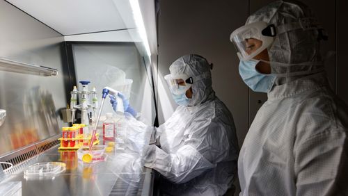
![[Translate to English:] Im GMP-Labor am Universitäts-Kinderspital soll die PV-Skin für den klinischen Einsatz hergestellt werden. Dabei gelten strengste Qualitäts- und Reinhaltevorschriften.](/fileadmin/_processed_/f/2/csm_03-Kunsthaut-003_S1A9843_ee6dca1bde.jpg)
Colour and capillaries
These two additional cell types are at the heart of PV-Skin therapeutics. Researchers led by Böttcher were able to add melanocytes and endothelial cells to denovoSkin, thus fabricating a skin substitute that contains four different cell types—a world’s first and yet another leap forward, as Böttcher relates. The pigment cells help restore the skin to its natural colour while also protecting it from UV rays, which is essential for healthy skin functioning. And the endothelial cells are involved in the formation of blood vessels—a sign indicative of faster, long-term healing.
But how is the skin substitute made? Put simply, the process consists of the following steps. First, a healthy skin sample the size of a postage stamp is removed from the patient. The sample is then dissolved in the lab using an enzyme solution and subsequently divided into four components: keratinocytes, fibroblasts, melanocytes and endothelial cells. Each cell type is placed in a separate Petri dish with a growth medium, where the cells replicate.
Afterwards, the individual components are mixed in a hydrogel and then reunited, as it were: first, a lower dermis layer consisting mainly of fibroblasts and endothelial cells is grown; then, the epidermis, primarily made up of keratinocytes and melanocytes, grows on top of the dermis. The actual process is naturally far more complex and sophisticated than this description suggests. “It’s like following a one-hundred-page recipe,” Sophie Böttcher says with a laugh.
Faster healing
So far, Böttcher’s team have fabricated the new PV-Skin in the labs at the Children’s Hospital and tested it in “test tubes” and on laboratory animals. The results are promising: the skin substitute is stable and capillaries form in the growth medium, enabling the artificial layer to attach itself to the underlying wound within just four days. “This boosts the supply of oxygen and the migration of cells, which brings about faster healing,” Sophie Böttcher says.
However, the planned clinical trials also increase the complexity of fabricating the skin substitute—which presents a problem at the hospital’s labs. “The official requirements placed on bioengineered developments are extremely strict, and in recent years, the number of specifications has grown exponentially, so that only a few labs in all of Europe will venture into clinical skin substitute research and production,” Böttcher says. The materials used in clinical care are also subject to the same high purity and quality standards required for production.

To comply with the regulations, the skin substitute must be fabricated in a GMP (Good Manufacturing Practice) lab where strict guidelines and directives determined and validated by the governmental authority responsible for drug approval (in Switzerland, Swissmedic) are in place. Sophie Böttcher says she and her team are currently working with the GMP lab at the Lausanne University Hospital to fabricate the skin substitute for the planned trials.
Lab as part of approval process
In the medium term, however, she hopes to relocate the production process to an in-house GMP lab at the University Children’s Hospital Zurich. The lab’s rooms have already been built in the research and teaching wing, located directly next to the acute care hospital. From the outside, it’s already obvious that the building is a high-security lab: some rooms have double-wall protection, while air pressure, humidity, temperature and particle count are continuously monitored. Researchers and technicians are even required to wear clothing designed to protect the products. Every single work step is predefined and carefully documented. Sophie Böttcher says the approval process could take another year yet.
Afterwards, the first patients can be treated in the Phase I clinical trials with the new PV-Skin. A key aim during this phase is demonstrating the product’s safety. Böttcher is confident of the outcome: “The preclinical trials gave no indication whatsoever that infections or side effects could occur.” What’s more, she’s convinced that the study is already delivering valuable data on the quality and benefits of the innovative skin substitute, all of which will aid the team in creating a stable, elastic and resilient skin substitute that’s as similar to natural skin as possible.
Facts and figures
Funding from the Werner Siemens Foundation
1.5 million Swiss francs
Project duration
2025 to 2028
Project leader
PD Sophie Böttcher, MD, Zentrum Kinderhaut, Brandverletzte Kinder, Plastische und Rekonstruktive Chirurgie, University Children’s Hospital Zurich
