Sophisticated skin substitute
Researchers at the University Children’s Hospital Zurich have developed the first-ever skin substitute to contain the four most important cell types found in natural skin. In a project recently awarded funding from the Werner Siemens Foundation, they’re now testing their innovation in clinical trials with patients.
Opened in November 2024, the new University Children’s Hospital Zurich is a breathtaking structure: with its arching lines, handsome wooden features and leafy courtyards, it certainly numbers among Europe’s most beautiful hospitals. The wards, clinics and operating theatres have been designed to maximise convenience, while the bright, modern patient rooms do their part to help children—and their parents—feel comfortable and well cared for.
Located on the building’s second floor is the centre for paediatric skin, where several hundred children and adolescents receive out-patient care for burns and scalding accidents every year. An additional one hundred and fifty children sustain such severe injuries that they require hospitalisation, with stays lasting from several days up to several months. Even after their burns have healed, many of these young patients need follow-up care in specialist consultations for paediatric burn victims, often until they reach adulthood.
Thin skin leads to thick scars
Significant advances have been made in therapies for severe burns, says privat-dozent Sophie Böttcher, deputy head of the hospital’s Division of Plastic and Reconstructive Surgery, Pediatric Burn Center. Despite the progress, however, limitations and challenges in burn treatment remain. For grave injuries, the standard approach is a skin transplantation using a patient’s own skin (an autograft). During this procedure, a thin layer of healthy skin—a split-thickness graft—is removed from the patient’s body and transplanted onto the sterilised wound.
Unfortunately, however, the method has several drawbacks. First, in cases of large-scale burns, there’s simply not enough intact skin left for taking a graft. Then, because the split-thickness graft is very thin, thick scar tissue often forms during the healing process, restricting movement and leaving unsightly marks. “Children with severe burns often suffer from the consequences of their injuries for the rest of their lives,” Böttcher relates.
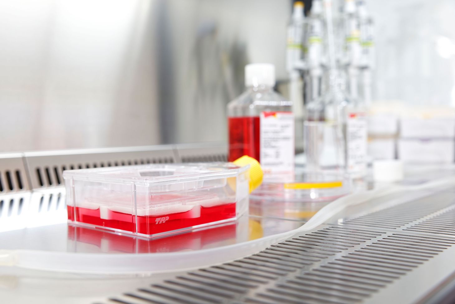
For more than a quarter century, clinicians and researchers at the University Children’s Hospital Zurich have been working towards creating a skin substitute using a patient’s own skin cells—work that was initiated and led by three professors at the time: Martin Meuli, Ernst Reichmann and Clemens Schiestl. The basic idea is to remove a healthy skin sample the size of a postage stamp from a patient, then isolate and replicate the individual cell types in the lab. Afterwards, the separate components are reunited, as it were, so they can develop into substitute skin tissue.
From two to four cell types
The first iteration of a lab-grown skin graft created at the hospital contains two key cell types found in skin: keratinocytes, the barrier-forming cells in the epidermis, and fibroblasts, which are found in the connective tissue of the dermis located beneath the epidermis. Compared to treatments with split-thickness grafts, this duo-cell skin substitute represents an enormous step forward, Sophie Böttcher says. “But in terms of quality, it’s nowhere near natural skin.”
By building on this earlier development, Böttcher and her team in the research group Skin and Soft Tissue Research Centre (SSTaRC) have now made considerable improvements to the approach. In addition to keratinocytes and fibroblasts, their new skin substitute—PV-Skin (P = Pigmented, V = Vascularised)—also contains two other vital skin cell types: melanocytes, or pigment cells, and endothelial cells, which are instrumental for the formation of blood vessels.
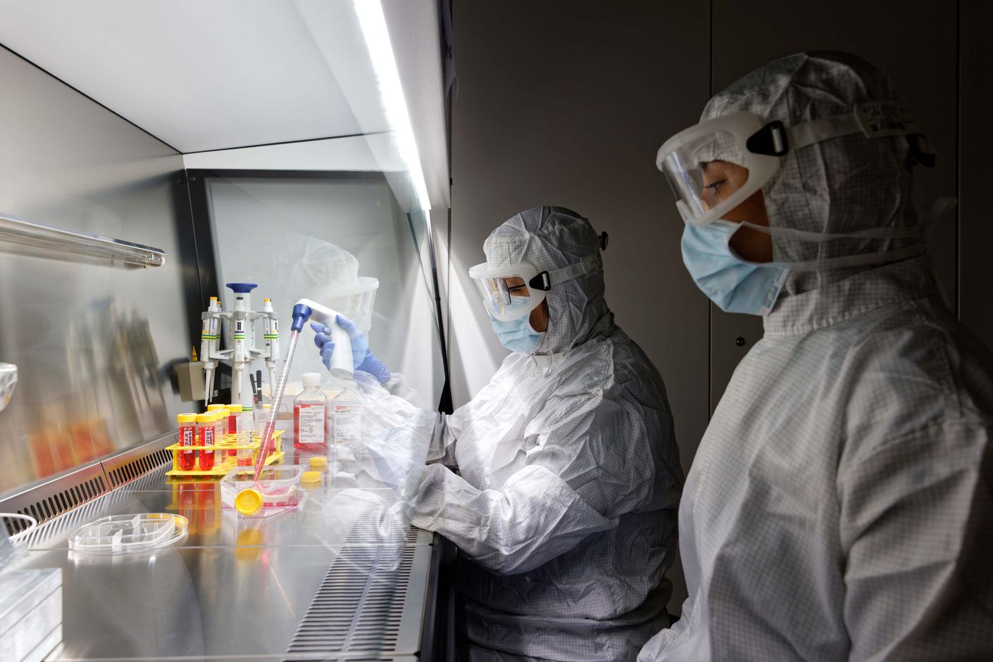
Laboratory and animal tests have delivered promising results, Sophie Böttcher says, adding that PV-Skin is astonishingly close to natural skin. Not only is it stable, but the presence of pigment cells ensures that the skin’s natural colour is retained. Moreover, the presence of endothelial cells promotes the fast development of capillaries, which in turn allow the skin substitute to attach itself to the underlying wound within just four days. “This boosts oxygen supply and the migration of cells, all of which brings about faster healing,” Böttcher explains.
The researchers are now ready to take the next step and conduct the first tests with PV-Skin on patients. Over the next three years, the clinical research team will fabricate and transplant skin grafts measuring seven-by-seven-centimetres for approximately ten patients. The Werner Siemens Foundation (WSS) is contributing a total of 1.5 million Swiss francs to the Phase I study, which has the primary aim of demonstrating the product’s safety and quality. In addition to Sophie Böttcher, patient care is also provided by privatdozent and clinical lecturer Dr. med. Kathrin Neuhaus, Chief Physician and Head of the Division of Plastic and Reconstructive Surgery, Pediatric Burn Center.
If the results meet the researchers’ expectations, more effective therapies may soon be available to children with burn injuries: the innovative skin substitute could greatly accelerate healing, reduce the need for follow-up surgeries and deliver a closer match to natural skin—ultimately improving the patients’ quality of life and promising a more hopeful future.