

Light-based bone fracture monitoring
How well a broken bone is healing can be determined by measuring blood flow and oxygen levels at the fracture site—as demonstrated in two studies in the “Smart implants” project that were led by WSS Endowed Chair Bergita Ganse. The new methods have the potential to enable simple, safe and seamless fracture monitoring.
Not all bone fractures heal well after surgery. “Complications occur in around fourteen of every one hundred tibial fractures—and affected patients often have to wait a long time before receiving a diagnosis,” says Bergita Ganse, WSS Endowed Chair for Innovative Implant Development at Saarland University.
In routine clinical practice, X-rays and CT scans are used to monitor bone fractures. However, frequent use of these imaging techniques is unsafe, as they expose patients to ionising radiation—a risk factor for cancer. In addition, these methods are unable to render protein structures visible; they only show the presence of calcium-based salts, which first form in a fracture gap at a late stage in the healing process. As Bergita Ganse explains, “This means it takes several weeks before these images can deliver information on how well a fracture is healing.”
Now, she and her team have developed a new method to enable more continuous monitoring of how well a broken bone is healing. Ganse says her previous work at the German Aerospace Center is what inspired the idea: “We measured blood flow and oxygen saturation in the muscles of astronauts—and I asked myself whether the same principle could be applied to measuring bones.”
New application for two methods
When a broken bone heals, numerous processes take place at the fracture site. “Filament-like structures form, which then begin to connect the ends of the fracture,” says Bergita Ganse. And gradually, new bone tissue supplied with blood develops. In addition, blood flow and oxygen saturation change, although how exactly this unfolds had never been studied in humans.
The practical devices currently on the market for measuring blood flow and oxygen saturation function on the basis of two different methods. First, laser-Doppler spectroscopy uses laser light to measure moving particles such as red blood cells—hence blood flow. And second, white light and near-infrared spectroscopy use LED light to assess a tissue’s properties, including oxygen saturation.
Together with Oana Scholz and Cedric Nowicki, two medical students in her team, Bergita Ganse used these standard devices to monitor fracture healing. Over the course of several months, they observed the healing process in a total of fifty-five patients with tibial fractures, then compared the findings with the results from a control group of fifty-one healthy individuals. In two recently published studies (*), the researchers demonstrate how blood flow and oxygen saturation change after a bone is fractured—and show that the healing timeline differs between fractures with a positive trajectory and those with suboptimal healing.
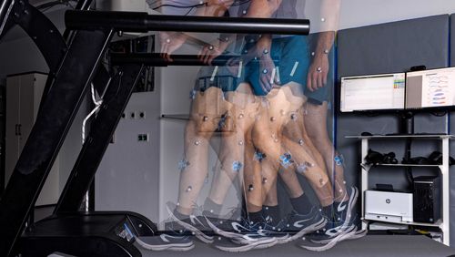
 first increases—followed by rising oxygen saturation levels (blue) once new blood vessels have formed.")

A characteristic image
Ganse says a characteristic pattern emerges after a bone fracture. “Blood flow initially increases to a maximum. Then, after about two to three weeks, the levels drop again.” Oxygen saturation, by contrast, first decreases to a minimum, and then rises again after two to three weeks, once new blood vessels have begun forming. “If these levels don’t return to normal, we have good reason to believe that healing isn’t progressing according to plan,” she says.
In future, doctors—or patients themselves—could place one of these measurement devices on the skin at the fracture site every day. After just a few seconds, and entirely without harmful radiation, they would know whether or not a fracture is healing well. “That’s not to say our method would completely replace X-rays,” Bergita Ganse says. “We’ll still need to see whether the screws are intact or whether a fracture gap has shifted. However, the new method means we can react faster when healing is suboptimal—and that’s extremely important.”
The sooner negative trajectories are detected, the better, especially because there are effective options for counteracting delayed healing. For instance, if a broken leg isn’t healing because a patient is walking too quickly or putting too much weight on the affected leg, doctors can prescribe more rest. Or if blood flow is insufficient—a common problem due to vascular damage in smokers or diabetics—the fracture site can be stimulated to increase blood flow. Possible therapies include ultrasound, shock wave therapy and magnetic field therapy.
Integration into the smart implant
Ideally, this function will one day be performed by the implant currently being developed in the WSS-funded “Smart implants” project, led by Bergita Ganse and Tim Pohlemann. The innovative device is designed to perform multiple tasks: stabilise a broken bone, provide data on how well a fracture is healing—and then react to this information. For example, if too much pressure is being applied to the fracture site, the implant will stiffen. And if patients don’t move enough, or blood flow at the fracture site is insufficient, the implant will change its shape, stimulating the healing process by giving the bone a kind of micro-massage.
Ganse says the blood flow and oxygen saturation measurements she and her team examined in the studies weren’t part of the original project idea, adding, “This makes it all the more exciting that we’re able to enhance the functionality of the implant.” She envisions integrating the oxygen saturation device into the implant itself. The advantage to this solution is that the device would be closer to the fracture site than when measurements are made through the skin—particularly relevant, given that the measuring depth of the devices is limited.
Comparing healing trajectories
Continuous monitoring of the healing process also enables the researchers to compare different treatment methods. “Until now, we could only compare the final products, as it were—meaning whether or not a fracture has healed properly,” Bergita Ganse explains. “But in future, we’ll be able to observe in real time how healing trajectories differ when a conventional implant or our smart implant is used.”
And there’s more. Ganse is fairly certain that the newly developed measurement method has the potential to bring benefits to other areas of bone health. In addition to lower-leg fractures, the system could be used to monitor other kinds of fractures as well as bone defects caused by tumours. “It will be extremely interesting to see how quickly the method can establish itself in research and in everyday clinical practice,” says Bergita Ganse.
(*)
Studie 1: https://doi.org/10.1016/j.bios.2025.117442
Studie 2: https://doi.org/10.3390/jfb15120384


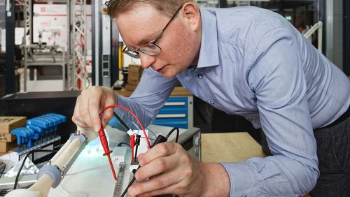
 in discussion with project leader Marcel Orth.")

![[Translate to English:]](/fileadmin/_processed_/c/a/csm_05_Miracle2_f0b59aff08.jpg "[Translate to English:]")