
Werner Siemens Imaging Center
Medical imaging techniques
The Werner Siemens Imaging Center plays in the premier league of research on medical imaging technology, with work on personalised tumour therapies forming part of Germany’s national Excellence Strategy. Now, thanks to new, combined medical imaging techniques, researchers are discovering which therapies work best for which patients.
Articles about the project

 is an international leader in the development of low-level radioactive tracers used in medical imaging.")


The project in brief
Despite major advances in modern medicine, certain diseases still evade detection and treatment – cancer and Parkinson’s to name just two. To remedy this, researchers at the Werner Siemens Imaging Center in Tübingen are developing and combining new imaging techniques that enable high-precision examination of tissues and molecules. The overall goal is to offer patients personalised therapies.
Cancer is the leading cause of early death in the western world. According to Bettina Weigelin, group leader at the Werner Siemens Imaging Center (WSIC) in Tübingen, Germany, the disease often returns after a few years, despite the good treatments available for many cancers. With her team, Weigelin is now seeking methods to better characterise tumours and to use this new knowledge to improve cancer therapies. Since 2021, the Tübingen researchers have benefited from a powerful new tool: their intravital microscope, an instrument that can be used to examine living tissue, even entire organisms. And this not just at the surface of tumour cells or other samples, but down to a tissue depth of two millimetres.
The group led by the centre’s director, Professor Bernd Pichler, is studying another phenomenon in tumour cells: senescence. Cells in a senescent state are still alive, but they no longer divide because they’re “sleeping”. Several cancer drugs exploit this mechanism to stop the growth of tumours. However, because it’s impossible to drive all of a tumour’s cells into a senescent state, and because senescent cells also secrete messenger substances that stimulate growth in the remaining cancer cells, it’s important to pinpoint exactly when the senescent cells should be destroyed with drugs. Ideally, this would prevent tumours that had already been suppressed from suddenly starting to grow again. To determine this precise moment, the researchers have developed a new procedure: a senescence tracer that can be used to detect senescent cells in the body. The team have already tested their method in preclinical trials. Currently, a phase I clinical trial is under way – the world’s first-ever clinical trial of a senescence tracer.
The WSIC team are also researching early detection of neurodegenerative diseases like Parkinson’s. Group leader Kristina Herfert and her team have developed the world’s first tracer to help doctors assess whether a therapy is working. After promising results in preclinical trials with mice and human brain tissue, the plan is to test the tracer already next year in a clinical trial with patients.
Facts and figures
Funding from the Werner Siemens Foundation
18.4 million euros (2024–2033)
15.6 million euros (2016–2023)
12.3 million euros (2007–2016)
Project leader
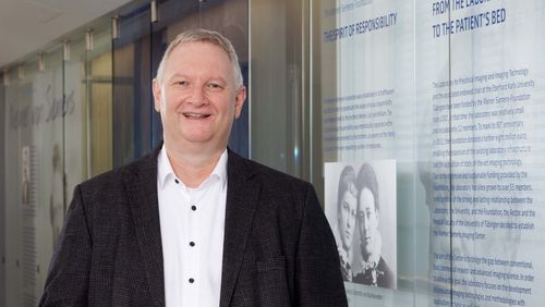
Prof. Dr Bernd Pichler, Werner Siemens Foundation Endowed Chair and Director of the Werner Siemens Imaging Center
Project duration
2007 to 2023