

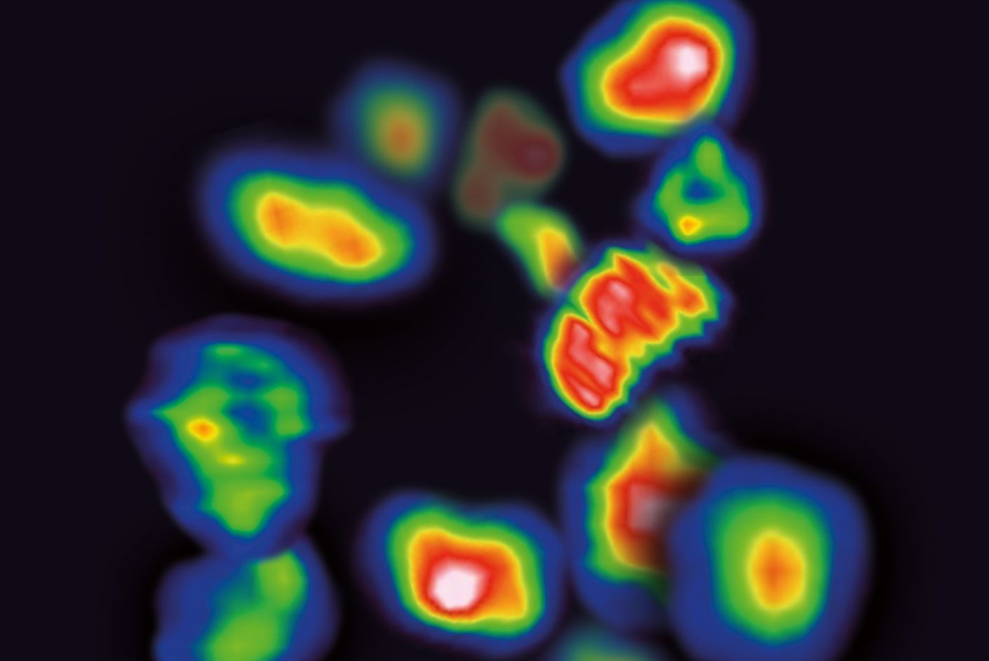
Using nanobodies in cancer diagnostics
Nanobodies—tiny fragments of antibodies—have the potential to fundamentally transform cancer diagnostics, and at the Werner Siemens Imaging Center (WSIC), researchers are using them to engineer highly specific PET tracers. Together with partners from research, clinical care and industry, the WSIC team are pursuing the ambitious aim of enabling future doctors to observe how a patient’s immune system responds to a therapy in real time.
The advent of immunotherapies that activate the body’s own ability to fight tumours has brought about a veritable sea change in modern cancer medicine, revolutionising how many forms of the disease are treated. Despite achieving some spectacular results, however, a core problem remains: in many patients, the therapies are either ineffective or they lose their efficacy over time.
“Immunotherapies are currently used to treat many kinds of tumours. But despite being very expensive, on average only twenty to thirty percent of all patients benefit from them,” says Dominik Sonanini, research group leader at the Werner Siemens Imaging Center (WSIC) at the University of Tübingen and specialist for internal medicine, haematology and oncology at the University Hospital Tübingen. “We want to know as early as possible which therapies are effective and when resistance to a therapy arises.”
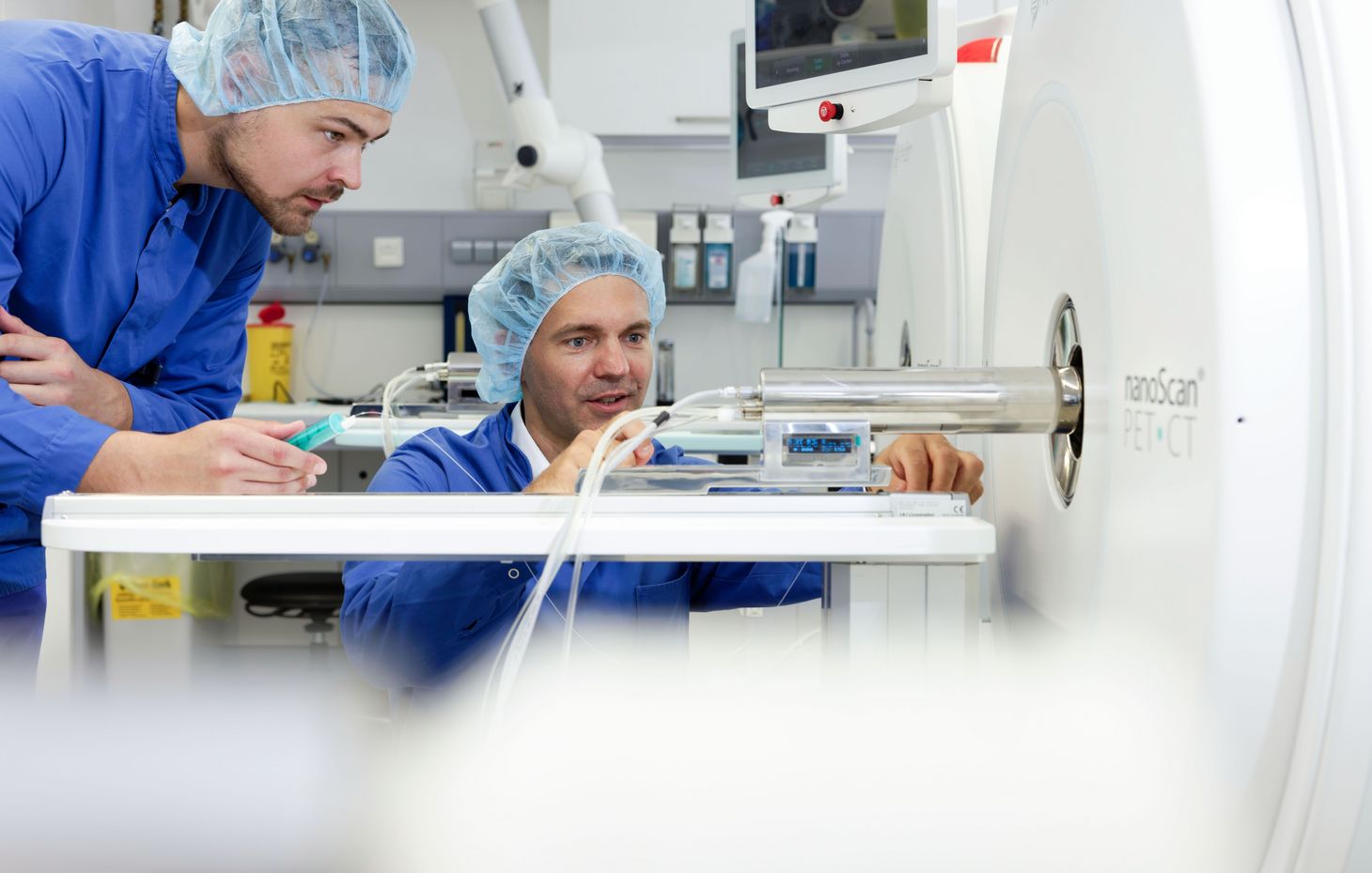
A promising instrument for detecting this information is molecular imaging of immune cells using positron emission tomography (PET). The team at WSIC are specialised in using radiolabelled substances, called tracers, to target different cell types of the immune system within the body. In addition to showing where immune cells are located, these markers also reveal how the immune cells change during treatment and shed light on the role they play in resistance mechanisms.
Six years for one nanobody
As part of their efforts towards achieving this aim, the WSIC researchers are focusing on one particular class of biological molecule: nanobodies. Nanobodies are the smallest, still functional antibody fragments that have been used in biomedical research to date. They were originally obtained from camelids such as alpacas, which possess a special type of antibody: the binding part of these molecules can be isolated and engineered.
By a stroke of luck, leading nanobody technology research groups are located just a stone’s throw from WSIC: the group led by Professor Ulrich Rothbauer at the University of Tübingen and researchers at the Natural and Medical Sciences Institute (NMI) in Reutlingen. Their proximity to WSIC has brought about a long-standing, fruitful partnership between the institutions—one that unites experience in the development and characterisation of nanobodies with expertise in tracer engineering and molecular imaging.
The road from alpaca molecule to sophisticated medical imaging technology is naturally long and complex. In a first step, the animals are immunised with the desired target molecule—a protein on the surface of immune cells, for example. Then, the researchers examine blood samples and identify nanobodies that bind to the specified target molecule. From the hundreds of possibilities that arise, they then select particularly stable and specific candidates that are suitable for a later use in humans. “It took three years to develop the first nanobody-based tracer, and another three years until we could fully validate it in animal models,” Sonanini says.
Antibodies in miniature
At approximately one tenth the length of a human antibody, nanobodies are small, to put it mildly. Their tiny size is, however, precisely what makes them so interesting. Because conventional antibodies remain in the circulatory system for several weeks, their presence interferes with medical imaging, as the constant background signals cover up the cell-specific signal.
“Unlike regular antibodies, nanobodies accumulate very quickly in tumour tissue, and anything that doesn’t form a bond is soon eliminated via the kidneys,” Sonanini explains. “As a result, we receive a highly specific signal with very little background noise.” This has several advantages. First, high-resolution images are generated already within an hour after the nanobodies are injected. Second, patients are exposed to less radiation. And third, even minimal changes are rendered visible. “We can see when the number of specific immune cells in a tumour has doubled,” Sonanini says.
Startup with a greater vision
The collaborative research activities led to the formation of the spin-off immuneAdvice in 2024. Under the leadership of CEO Teresa Wagner, the company’s mission is transferring preclinical developments into clinical care.It should, however, be noted that the vision at immuneAdvice involves much more than one single tracer—the company has plans to develop an entire platform of nanobody-based imaging molecules.
“Our idea is to create a pipeline with tracers targeting different immune cells and immune checkpoints,” says Sonanini, who also acts as immuneAdvice’s Chief Medical Officer. The multipronged approach addresses the fact that an immune response is never determined by a single cell type, as T cells, natural killer cells, macrophages and regulatory immune cells all engage in complex interactions. In order to understand why a therapy is effective or why it fails, it’s important to simultaneously consider as many of these factors as possible.
A long-term aim at immuneAdvice is to chart the immune system, creating a type of map. For this, several tracers could be used in succession to generate a comprehensive image of the immune activity in a patient within a short period of time. “Just one piece of information will never be enough” Sonanini explains. “In future, the combination of various data and their AI-assisted interpretation will form the basis for decisions on personalised therapies.”
Despite the advances made, the path to clinical care is far from straightforward. Nanobodies are biological proteins and as such are subject to strictly regulated good manufacturing practices (GMPs). Together with the NMI Biological Development Center and with financing from the European Regional Development Fund, a centre of excellence was created with the aim of accelerating these processes and to make them available for partners in academia and industry. Nevertheless, developing a clinically viable product costs millions of euros. The high costs are another reason for creating immuneAdvice: to attract investors and secure financing for the complex approval processes. The venture is already paying off: Sonanini says immuneAdvice has recently acquired 1.6 million euros in seed-funding.
Looking inside a tumour
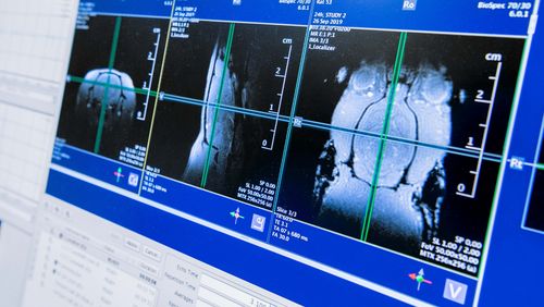
In a study published last year in top-tier journal Science Advances (1), the team demonstrated the great potential held by nanobody-based tracers. They developed a PET tracer for targeting the CD4 receptor, a surface marker on T helper cells, enabling them to produce the first-ever visualisation of the spatial distribution of these immune cells on the inside of a tumour.
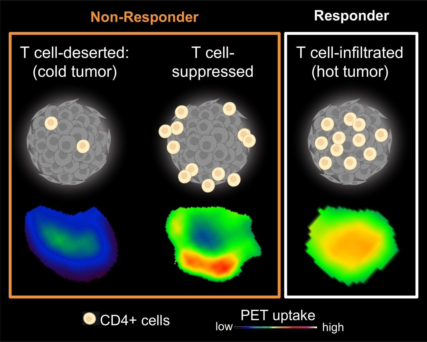
The researchers were then able to observe that successful immunotherapies were linked to CD4 cells migrating to the core of a tumour. If, however, the cells remained largely on a tumour’s surface, the therapy was significantly less effective. Sonanini says that, in animal models, these patterns enabled the effectiveness of a treatment to be predicted just a few days after a therapy began: “We not only detected whether the cells responded to the tumour; we were also able to draw conclusions about certain resistance-related mechanisms.”
Sonanini also believes the results illustrate the added value nanobody-based radiotracers can bring to clinical care. “With today’s methods, it can take months before we see that a therapy isn’t working,” he says. “With nanobody-based tracers, we think we’ll need just four to six weeks to determine whether an adjustment is necessary.”
Towards precision medicine
The published study also exemplifies the constructive research culture practised in Tübingen. The nanobody experts at NMI and the University of Tübingen, the imaging specialists at WSIC and the Department of Nuclear Medicine, the team in radiopharmacy and immuneAdvice all collaborate closely. “The excellent teamwork and dedication of all involved are driving the translation of an idea from basic research into clinical care,” Sonanini says. “The long-term funding provided by the Werner Siemens Foundation lays the basis for realising a project of this magnitude at WSIC.”
Although the technology is still at an early stage, the way forward is clear: developing a state-of-the-art imaging technology that’s capable of visualising every aspect of the human immune system associated with fighting tumours. The nanobodies developed in Tübingen promise to deliver a valuable tool that helps physicians to select the right therapies, identify resistance earlier, and customise a treatment to an individual patient’s needs.
Text: Simon Koechlin
Translation: Mary Carozza
> Study