

Detecting Parkinson’s-related protein in the brain
Eine Publikation des Werner Siemens Imaging Centers weckt Hoffnung für die frühzeitige Diagnose und individuelle Behandlung von Parkinson und verwandten Erkrankungen: Das Forschungsteam unter der Leitung von Kristina Herfert hat einen neuartigen PET-Tracer entwickelt, der krankhafte Ablagerungen des Proteins Alpha-Synuclein im lebenden Gehirn sichtbar macht.
Seit Jahren suchen Forschende nach einer Möglichkeit, die eigentliche Ursache von Parkinson und verwandten Erkrankungen direkt im lebenden Gehirn sichtbar zu machen. Nun hat ein Team um Prof. Kristina Herfert vom Werner Siemens Imaging Center (WSIC) an der Universität Tübingen, Prof. Christian Griesinger, Direktor am Max-Planck-Institut für Multidisziplinäre Naturwissenschaften und Prof. Armin Giese, Chief Scientific Officer des Biotechnologie-Unternehmens MODAG einen wichtigen Schritt zu diesem Ziel gemacht: In der renommierten Fachzeitschrift «Science Translational Medicine» stellen die Forschenden einen PET-Tracer vor, mit dessen Hilfe sie krankhafte Ablagerungen des Proteins Alpha-Synuclein in Hirngewebe, Tiermodellen und erstmals auch bei Patienten nachweisen.
Alpha-Synuclein ist kein Fremdkörper im Gehirn. Das kleine Eiweiss erfüllt dort eine wichtige Funktion bei der Signalübertragung zwischen Nervenzellen. «Die Kommunikation zwischen Nervenzellen funktioniert über sogenannte Neurotransmitter», erklärt Kristina Herfert. Alpha-Synuclein sei an diesem Prozess beteiligt. Problematisch wird es, wenn sich das Protein falsch faltet. Dann entstehen zunächst kleine Aggregate und später grössere Ablagerungen, die als Kennzeichen einer Gruppe von neurodegenerativen Erkrankungen gelten. Dazu gehören die Parkinson-Krankheit, die Lewy-Körper-Demenz und die Multiple Systematrophie (MSA).

An article co-authored by scientists at the Werner Siemens Imaging Center is raising new hope for the early diagnosis and personalised treatment of Parkinson’s and other related diseases. Under the leadership of Kristina Herfert, a research team developed a PET tracer for detecting accumulations of abnormal deposits of the alpha-synuclein protein in the brains of living patients.
Researchers have long been seeking a way to visualise the underlying cause of Parkinson’s and related diseases directly in the brains of living patients. Recently, a team led by Kristina Herfert (Werner Siemens Imaging Center WSIC, University of Tübingen), Christian Griesinger (Director at the Max Planck Institute for Multidisciplinary Sciences) and Armin Giese (Chief Scientific Officer at biotechnology company MODAG) have taken a significant step towards this aim by developing a positron emission tomography (PET) tracer that can detect abnormal alpha-synuclein deposits in human brain tissue, animal models—and even living patients. They introduced their potentially groundbreaking tracer in an article published by top-tier journal Science Translational Medicine.
Far from being a foreign substance in the brain, alpha-synuclein plays a vital role in the transfer of information from one nerve cell to another. “Communication between nerve cells functions via neurotransmitters,” Kristina Herfert explains, adding that alpha-synuclein is involved in this process. Problems arise when the protein misfolds, which first causes small aggregates, then larger deposits of abnormal alpha-synuclein to form. These accumulations are the main attribute in a family of neurodegenerative diseases that includes Parkinson’s disease, Lewy body dementia and multiple system atrophy (MSA).
More difficult to detect than Alzheimer’s
The precise role these aggregates play is still not fully understood. “Many scientists believe it’s mainly the early forms of aggregation that are toxic and responsible for cell damage,” Herfert says. This hypothesis explains why researchers are interested in intervening in the process at a very early stage—and why the ability to detect alpha-synuclein deposits as soon as possible is needed for such interventions.
Although imaging techniques for visualising the amyloid plaques formed in Alzheimer’s have been in use for many years now, an instrument for detecting the similar alpha-synuclein aggregates that arise in Parkinson’s has thus far remained elusive. Herfert cites numerous reasons for this difficulty, one being that there are considerably fewer abnormal alpha-synuclein deposits in Parkinson’s than amyloid plaques in Alzheimer’s.
Another reason is that the Parkinson’s-related aggregates are located inside nerve cells. “In addition to penetrating the blood-brain barrier, a tracer molecule for detecting these aggregates also has to go through the cell wall,” Herfert says. “This makes the entire process much more complicated.” In addition, the alpha-synuclein structure resembles that of other disease-relevant proteins such as tau or amyloid beta proteins. Consequently, a method must be highly selective in order to conclusively detect Parkinson's.
Results after twelve years
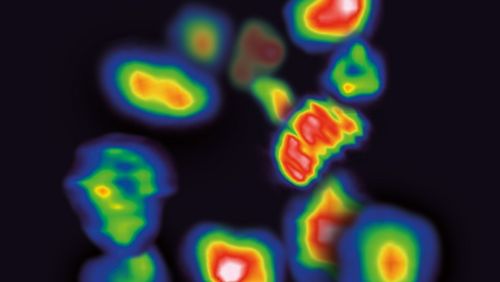
One of the most important tools for detecting metabolic processes and products in the body is tracer-based imaging using PET technology. Tracers are substances with very low levels of radioactivity that are injected into the body, where they accumulate on or in specific cell types. A common radioactive marker is the carbon isotope 11C. A PET scanner recognises the marker, thereby visualising metabolic processes as well as lesions such as alpha-synuclein deposits in brain cells.
The official name of the new PET tracer is [¹¹C]MODAG-005, and its origins date back to 2013 and the European Union research project MultiSyn, an international undertaking coordinated by the University of Tübingen in which the biotech firm MODAG was one of the participants. “Twelve years of research were needed before we could complete and test the current version of the molecule in 2025,” Herfert says. She also relates that numerous setbacks were encountered along the way. For instance, a promising early version of the molecule, MODAG-001, demonstrated a proclivity for binding with alpha-synuclein. However, it also simultaneously formed non-specific bonds with other proteins in the brain tissue, which caused quite a bit of background noise and greatly reduced the tracer’s signal. To resolve this, the researchers altered one specific chemical group in the molecule. “This modification led to a significantly better signal,” Herfert says.


Tests in the lab and with patients
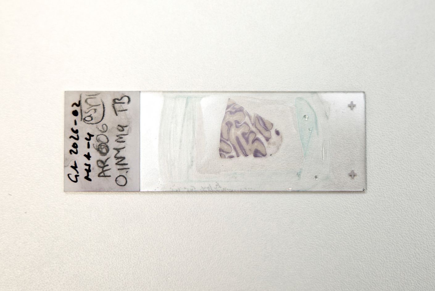
For their publication, the researchers tested their new tracer in various settings. They first worked with synthetic alpha-synuclein fibrils, then with brain tissue from deceased patients. MODAG-005 demonstrated an excellent binding affinity with the abnormal protein deposits. At the same time, the researchers also observed that their molecule demonstrated a much greater affinity for alpha-synuclein than for tau proteins or amyloid deposits.
Following these lab tests, the researchers turned to animal experiments with mice, rats and non-human primates. When alpha-synuclein fibrils were injected into the brains of rodents, the tracer gathered specifically in the affected regions of the brain. Increased uptake was also detected in heavily affected areas in the brains of genetically altered Parkinson’s mice.
After attaining these positive results, the researchers were ready to conduct a small study with humans. They evaluated two patients with MSA, one patient with Parkinson’s and one control person showing no symptoms of a synuclein-related disease. The findings revealed that alpha-synuclein aggregations were indeed detected in the patients with MSA and Parkinson’s. Even more interestingly, the distribution of the tracers in those patients corresponded to the different clinical manifestations of their respective diseases.
In short, the PET scans showed precisely those patterns that were expected based on the known progression of the diseases. For instance, strong signals were visible in the cerebellum and brain stem of the MSA patients, whereas the clearest signals appeared in the basal ganglia of the Parkinson’s patient. Only weak signals were detected in the control person. “In this test, our tracer differentiated between the two neurodegenerative diseases,” Herfert says.
More than just a diagnostic tool
It should also be noted that the tracer’s true value goes beyond its ability to establish a differential diagnosis. To date, Parkinson’s research has lacked a direct biomarker for controlling whether a medication has the desired effect on abnormal protein deposits. MODAG-005 has the potential to break new ground in this area, as the study also revealed that the experimental drug Emrusolmin developed by MODAG inhibits the tracer. It appears that both molecules bind to the same alpha-synuclein aggregate structures.
Despite the excellent results, however, the study authors caution against drawing premature conclusions. The test setting with patients involved only three individuals with different but pronounced clinical presentations. It remains to be seen whether MODAG-005 is sensitive enough to detect the diseases before distinct symptoms appear. Herfert reiterates that the patients examined had already been given a clear diagnosis, adding, “It will be interesting to see whether the tracer can identify aggregates earlier in the progression of the disease.”
Herfert says she’s very optimistic about the binding affinity of MODAG-005. It’s located in the subnanomolar range, one of the best affinities that have been specified for alpha-synuclein tracers. Currently, the researchers are preparing for the start of a Phase I clinical trial with their novel tracer. If the previous results are verified, MODAG-005 could become a vital instrument in Parkinson’s research—similar to the amyloid PET scans that have changed Alzheimer’s research over the past two decades. For patients, this would represent a key step towards receiving early-stage, personalised therapies.
Text: Simon Koechlin
Translation: Mary Carozza
> Studie